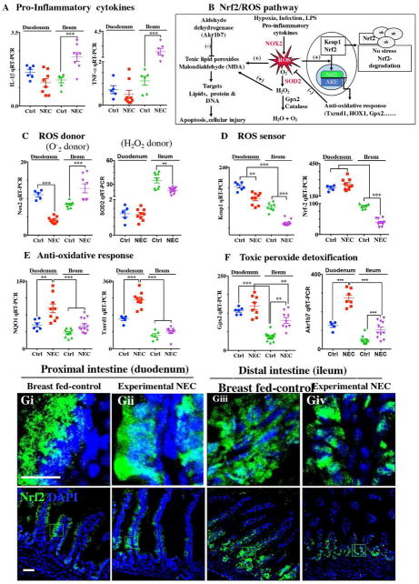
Figure 8. The anti-oxidative environment of the proximal small intestine versus the distal small intestine correlates with the location of NEC.
A: qRT-PCR of pro-inflammatory cytokines IL-1b and TNF-a; B: Schematic of Nrf2/ROS oxidative injury pathway in intestinal epithelium; C: qRT-PCR of ROS donor enzymes Nox2 and SOD2; D: qRT-PR of ROS sensor Keap1 and Nrf2; E: qRT-PCR of antioxidants NQO1and Txnrd1; F: qRT-PCR of peroxide detoxifier Gpx2 and Akr1b7; G: Immunofluorescence images of Nrf2 showing cytoplasmic and nuclear translocation (Nrf2-green) and DAPI (nuclei, blue) in control and NEC mice (Ctrl, Control-Breast fed or experimental NEC. **p<0.01, ***p<0.001 by a Student t test when comparisons of two groups were made, and by ANOVA for multiple comparisons, each dot represents data from individual mouse. ROS (reactive oxygen species), Nox2 (NADPH oxidase), SOD2 (Superoxide dismutase, mitochondrial), Keap1 (Kelch Like ECH Associated Protein 1), Nrf2 (NF-E2 p45-related factor), NQO1 (NADPH Quinone Dehydrogenase 1), Txnrd1 (Thioredoxin Reductase 1), Gpx2 (Glutathione Peroxidase 2, intestinal), Akr1b7 (Aldo-keto reductase family 1, member B7), scale bar =10μm.

