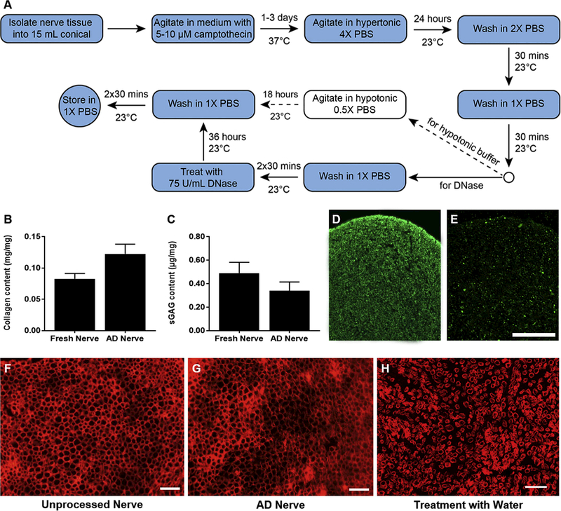
Figure 3:
Apoptosis decellularization preserves desirable matrix components and structure but not myelin proteins. (A) Flow diagram of the generalize process for apoptosis decellularization. Dashed arrows indicate steps that were excluded from the final process for nerve tissue. (B) Quantification of collagen content before and after apoptosis decellularization (n=4). (C) Quantification of sulfated GAGs before and after apoptosis decellularization (AD) (n=5). Neither quantification revealed a significant difference. Immunoreactivity for myelin basic protein was assessed for unprocessed nerve (D) and apoptosis decellularized nerve (E). (F-H) Fluorescence micrographs of laminin staining in fresh nerve (F), apoptosis decellularized nerve (G), and fresh nerve processed with water for cell lysis (H). Processing and staining was repeated at least three times per group. Scale bars represents 50 μm. No significant difference was found in B or C.

