Figure 5:
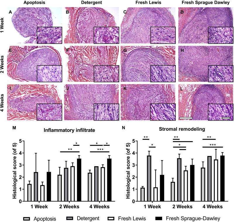
Apoptosis-decelluarized nerves are immunologically tolerated, with decreased inflammatory infiltrate and stromal remodeling compared to isograft by 4 weeks. Tissue from one of four groups (apoptosis decellularized, detergent decellularized, or fresh Lewis and Sprague-Dawley controls) was implanted subcutaneously for 1–4 weeks (n=4). (A-L) Brightfield H&E images for an example explant per group showing areas of infiltrates and stromal changes at weeks 1, 2, and 4. Images are at 5x with 40x inset images to show detail within the implant. Scale bars represent 500 μm for 5X images and 50 μm for 40X images. (M) Quantitative histological scoring of inflammatory infiltrates at the center at 1, 2, and 4 weeks. (N) Quantitative scoring of stromal cells at the center at 1, 2, and 4 weeks. Error bars represent standard error with n=4 except detergent decellularized at 4 weeks (n=1). *p<0.05 **p<0.01 ***p<0.001.

