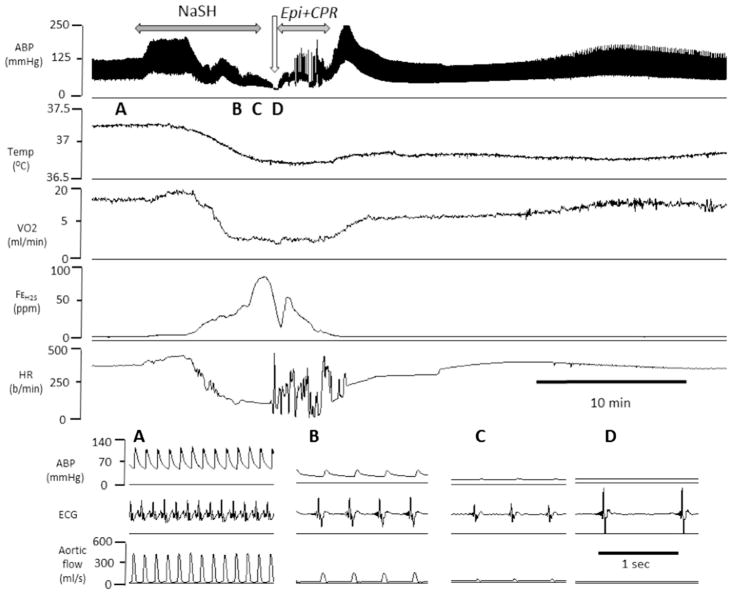
Fig. 10.
Example of the effects of epinephrine after H2S infusion (0.8 mg/min)-induced PEA in one rat. From top to bottom, carotid blood pressure (ABP), temperature, V̇O2, expired fraction of H2S and HR are displayed. During sulfide infusion, blood pressure as well as V̇O2 decreased leading to a typical PEA as shown in the insets A–D. Injection of epinephrine during complete cardiac asystole followed by chest compressions allowed ROSC

