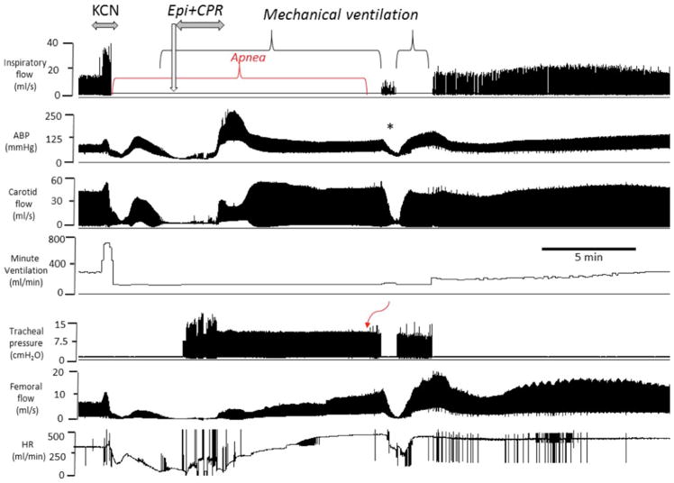
Fig. 8.
Example of the effects of epinephrine following KCN infusion (0.75 mg/kg/min) induced PEA in another spontaneously breathing rat. From top to bottom, inspiratory flow, arterial blood pressure, carotid blood flow, minute ventilation, tracheal pressure, femoral flow and hear rate are displayed. Like in Fig. 7, following a brief period of hyperventilation, an apnea occurred followed by a rapid decrease in blood pressure, blood flow and heart rate leading to a PEA. Epinephrine was administered while mechanical ventilation was initiated. Circulation was restored after the injection of epinephrine with chest compressions. The asterisk (*) shows a period where mechanical ventilation was briefly interrupted while breathing was present but still depressed, which was associated with a drop in blood pressure and blood flow. Note that spontaneous breathing movements started to be generated one minute later in a stable manner

