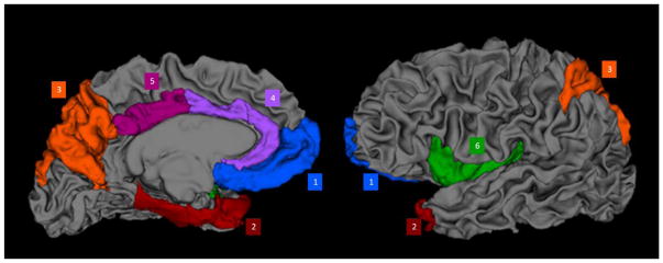
Figure 1.
A 3-dimensional representation of the brain’s white matter derived in FreeSurfer with the cortical areas used in the study superimposed and labeled by color. Blue = medial prefrontal lobe (medial orbital and frontal pole); Red = temporal (entorhinal cortex, parahippocampus, temporal pole); Orange = parietal (superior parietal lobule, precuneus); purple = anterior cingulate cortex (caudal and rostral anterior cingulate cortex); magenta = posterior cingulate cortex; green = insula.

