
Figure 1.
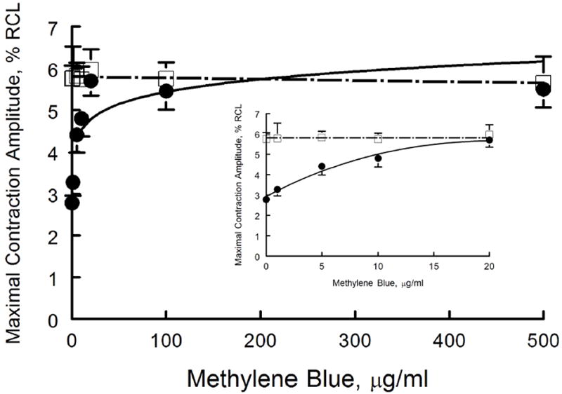
MB rescues contractile dysfunction of cardiomyocytes exposed to H2S: MB dose response. Freshly isolated myocytes from mouse LV and septum were plated on laminin-coated coverslips, bathed in medium 199 ([Ca2+]o 1.8 mM) and paced (2 Hz) to contract (37°C)(Methods). MB (0 to 500 μg/ml) or NaHS (100 μM) + MB (0 to 500 μg/ml) were added at time 0, and contractions were measured at 10 min. Maximal contraction amplitudes (% of resting cell length, %RCL) are shown for MB (□) and NaHS + MB (●) myocytes. For MB alone, there were 20, 7, 10, 6, 10, 8 and 6 myocytes at 0, 1, 5, 10, 20, 100 and 500 μg/ml, respectively. For NaHS + MB, there were 25, 8, 9, 8, 9, 7, and 8 myocytes at 0, 1, 5, 10, 20, 100 and 500 μg/ml of MB, respectively. Inset: expanded view of contractile response at MB doses from 0 to 20 μg/ml.