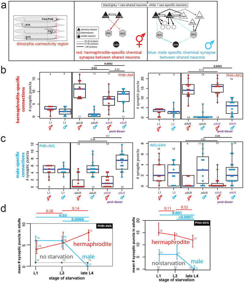
Fig.1: Starvation inhibits male-specific synaptic pruning.
(a) Schematic of adult chemical synaptic connectivity based on electron micrograph reconstruction3,4. Left panel shows location of dimorphic synaptic connections within animal. mMN = male motor neuron, mIN= male interneuron.
(b) Hermaphrodite-specific PHB>AVA and PHA>AVG synaptic connections are maintained in post-dauer adult males as quantified by GRASP and iBLINC, respectively. Each dot represents one animal (red=hermaphrodite, cyan=male in all figures, n=number of animals, shown in each column), blue bars show median, black boxes represent quartiles, vertical black lines show range (b,c,d). Control animals are the progeny of the post-dauer animals. L1 animals (pre-synaptic pruning) shown to the left. p-values shown by two-sided Wilcoxon rank-sum test with Bonferroni corrections for multiple testing (where applicable; see Methods) (b,c,d). Representative images shown Extended Data Fig.1a. While we observed a decrease in synaptic puncta in starved PHB>AVA hermaphrodites, this was not consistently reproducible across experimental replicates and thus is likely an experimental artifact.
(c) The male-specific PHB>AVG and AVG>DA9 synaptic connections prune normally in post-dauer adult hermaphrodites. Representative images shown Extended Data Fig.1a.
(d) Starvation in the L1 or L3 stages, but not L4, results in a failure of PHB>AVA and PHA>AVG to prune in males. Animals were starved for 24 hours (12 hours of starvation was insufficient to affect male-specific pruning). Center is mean number of synaptic puncta, error bars show standard deviation. “No starvation” shows progeny of starved animals as control. n=number of animals, shown above each data point.

