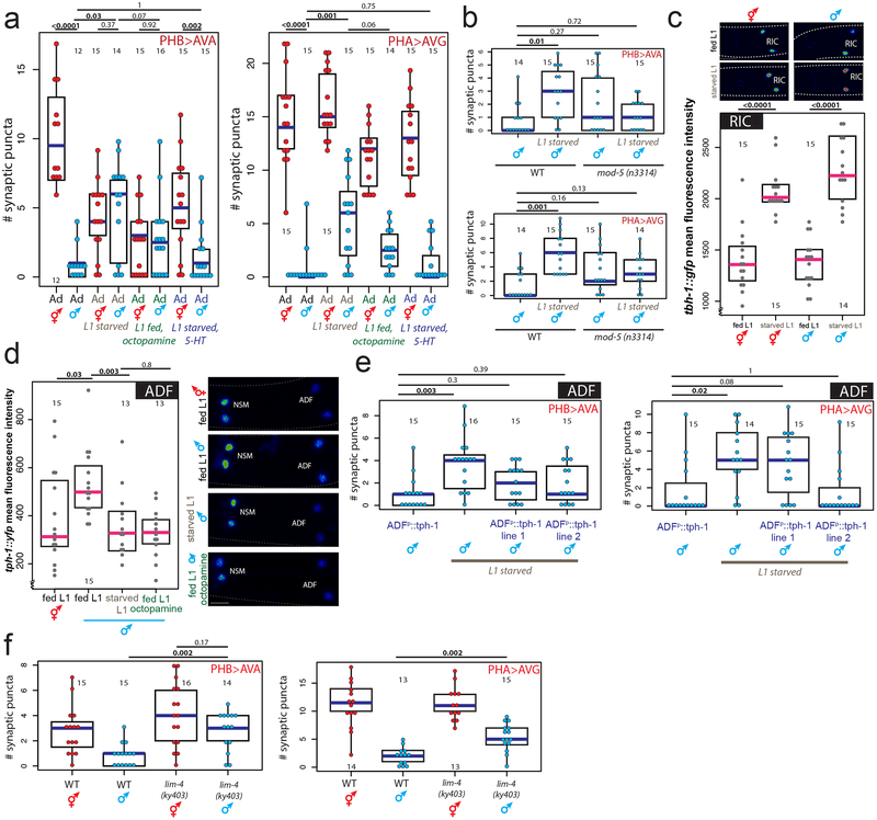
Fig.3: Serotonin and octopamine convey feeding and starvation signals via the ADF neurons.
(a) Octopamine and 5-HT (serotonin) mimic starvation and feeding, respectively. Quantification of PHB>AVA and PHA>AVG synaptic connectivity in adults of both sexes using GRASP or iBLINC (a, b, e, f).. Each dot represents the number of synaptic puncta in one animal, blue bars median, black boxes quartiles, vertical black lines range (a, b, e, f). n=number of animals, shown in each column (a, b, c, d, e, f). p-values shown by two-sided Wilcoxon rank-sum test with Bonferroni corrections (where applicable; see Methods) for multiple testing (a, b, c, d, e, f). Representative images shown Extended Data Fig.3a.
(b) mod-5 mutants rescue pruning defects in PHB>AVA and PHA>AVG connections in L1-starved adult males. Representative images shown Extended Data Fig.3c.
(c) Starvation induces octopamine production. Expression levels of a tbh-1 reporter in RIC in fed or starved L1 animals. Heat-map rendered fluorescence intensity images above, quantification below. Scale bars, 10μm, all panels. Magenta bar median, black boxes quartiles (c,d). Anterior left, dorsal up in all figures.
(d) tph-1 transcription in the ADF neurons is increased in males and decreased by starvation or exogenous octopamine. Expression levels of a tph-1 transcriptional fosmid in ADF in fed L1 animals, starved L1 animals, or L1 animals fed in the presence exogenous octopamine.
(e) tph-1 overexpression in ADF mimics the rescuing effect of exogenous 5HT. Quantification of PHB>AVA and PHA>AVG synaptic connectivity using GRASP and iBLINC. Two independent transgenic lines were tested for each experiment, L1 starved animals without transgenic lines are siblings of transgenic animals, controls are non-starved adult males with transgenic arrays. Representative images shown Extended Data Fig.6c.
(f) Genetic ablation of ADF results in failure to prune. Quantification of PHB>AVA and PHA>AVG synaptic connectivity in both sexes in lim-4 mutants24.

