
FIG. 3.
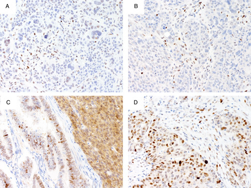
(A, B) Endometrial serous carcinoma with complete absence pattern of abnormal p53 expression stained on 2 different platforms. (A) Nonspecific nuclear staining interpreted as wild-type pattern; (B) shows complete absence of nuclear staining but a weak cytoplasmic blush indicating staining bordering on too strong. (C) Endometrial endometrioid carcinoma with wild-type staining with slight cytoplasmic blush on the left and true abnormal cytoplasmic staining on the right (compare with low-power view in Fig. 4C). The true abnormal cytoplasmic staining is accompanied by a variable nuclear staining of similar intensity but not strong diffuse. (D) Endometrial endometrioid carcinoma with wild-type pattern showing weak cytoplasmic staining probably due to too strong staining. This should not be interpreted as abnormal cytoplasmic staining.