Abstract
The study was under taken to know the prevalence of reflux signs in an individuals with throat complaints on the basis of reflux finding score (RFS) and quantitatively assess the effect of treatment. A cross-sectional study was done to evaluate the presence of laryngo-pharyngeal reflux signs in patients visiting ENT clinic with throat or voice problems in central India. There were 80 patients included in the study from 2017 to 2018 individuals. They were questioned regarding their symptoms. Their pharyngeal findings on rigid 70° laryngoscopy were viewed and RFS was made. The patients were reviewed at monthly intervals. Laryngopharyngeal reflux changes were seen in 36 of the 80 patients (45%). The reflux was graded as per the reflux finding score. The score ranged from 7 to maximum of 17 out of 26 in the patients with LPRD. Majority of the patients the score decreased with lifestyle changes and pantaprazole twice daily. There was poor response in 5% (4) patients, who were then advised to undergo upper gastro intestinal endoscopy for further assessment. Laryngopharyngeal reflux has become a very common entity in urban lifestyle. On careful examination the signs can be picked and assessed with the RFS, which is a very useful tool to grade and reassess patient on subsequent follow up.
Keywords: Laryngopharyngeal reflux, Reflux finding score, Lifestyle modification, Proton pump inhibitors
Introduction
The change in eating habits and sedentary life style, increased stress, tight clothing, desk jobs has led to a rise in patients with reflux. Reflux is defined as a return of fluid. Reflux can be of the gastric contents into the oesophagus or more upwards into the larynx and pharynx. Reflux of gastric content into larynx and pharynx is known as laryngopharyngeal reflux disease (LPR).
According to the Montreal Consensus Conference, the manifestations of gastroesophageal reflux disease (GERD) have been classified into either esophageal or extraesophageal syndromes and, among the latter ones, the existence of an association between LPR and GERD has been established Vakil et al. [1] There is difference in the symptomatology of GERD and LPR. The patterns, mechanisms, manifestations, and treatment of laryngopharyngeal reflux (LPR) and gastroesophageal reflux disease (GERD) differ, and the gastroenterology model of reflux disease does not apply to LPR. LPR patients have head and neck symptoms, but heartburn is uncommon. Consequently, LPR is often called silent reflux. LPR patients have predominantly upright (day-time) reflux and normal esophageal motility; most do not have esophagitis, which is the diagnostic sine qua non of GERD. Moreover, the laryngopharyngeal epithelium is far more susceptible to reflux-related tissue injury than is the esophageal epithelium. Because of these differences, treatment algorithms for LPR and GERD vary [2].
Materials and Methods
This is a cross sectional study done in patients presenting to the otolaryngologist for throat problems of more than 1 month duration. The prevalence of LPRD was evaluated on the basis of reflux Finding score (RFS) and the effect of treatment was re-evaluated on the basis of RFS (Table 1). Acute illness were excluded from the study. There were 80 patients in the study group. The patients were followed up at monthly intervals. A through ENT examination was done followed by rigid 70° laryngoscopy.
Table 1.
Reflux finding score.
Adapted from Belafsky et al. [16]
| Findings | Score |
|---|---|
| Subglottic edema | 0 = absent 2 = present |
| Ventricular obliteration | 2 = partial 4 = complete |
| Erythema/hyperemia | 2 = arytenoids only 4 = diffuse |
| Vocal fold edema | 1 = mild 2 = moderate 3 = severe 4 = polypoid |
| Diffuse laryngeal edema | 1 = mild 2 = moderate 3 = severe 4 = obstructing |
| Posterior commissure hypertrophy | 1 = mild 2 = moderate 3 = severe 4 = obstructing |
| Granuloma/granulation of tissue | 0 = absent 2 = present |
| Thick endolaryngeal mucus | 0 = absent 2 = present |
Observations
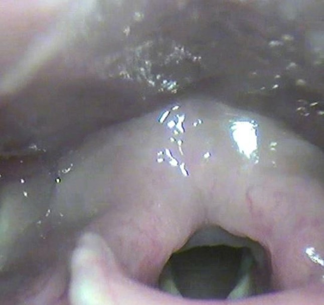
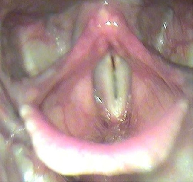
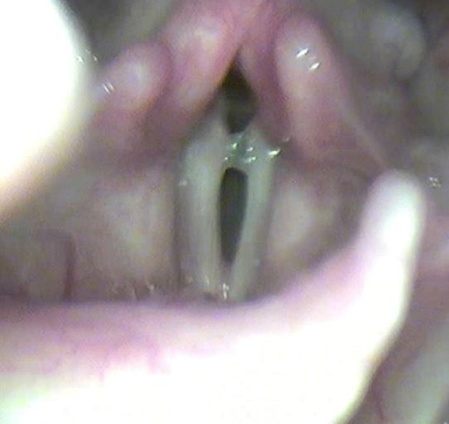
In the study group the prevalence of LPRD was 45% (RFS more than 7) in patients with throat complaints (Table 2). The maximum RFS was 17. Patients were commonly found to have posterior commissure hypertrophy (Fig. 1), hyperaemia (Fig. 2). Patients with thick endo-laryngeal mucus (Fig. 3) had complaints of voice change and constant throat clearing. The remaining patients with low RFS were found to have other causes like post nasal drip and allergy as the cause for throat irritation. The patients with high RFS were given proton pump inhibitors twice daily along with life style modifications. The patients with low RFS with no other identifiable cause were given lifestyle modification. These patients were followed up at 1 and 2 month duration. Majority were found to symptomatically improve in 15 days with decrease in RFS on the monthly follow up (Table 3). Four patients (5%) did not show significant improvement were advised to undergo upper gastrointestinal endoscopy. Proton pump inhibitor were given for 2 months. Patients were asked to modify lifestyle and dietary habits. However these patients tend to relapse if the lifestyle modification is not continued indicating a genetic/anatomic basis for LPR in certain individuals who are more prone to reflux.
Table 2.
Reflux finding score at diagnosis
| Reflux finding score | Number of patients (n = 80) |
|---|---|
| Less than 7 | 44 |
| More than 7 | 36 |
Fig. 1.

Reflux finding score 12—posterior commissure hypertrophy
Fig. 2.

Reflux finding score 8—hyperemia
Fig. 3.

Reflux finding score 7—thick endolaryngeal mucus
Table 3.
Reflux finding score at 2 months
| Reflux finding score | Number of patients (n = 80) |
|---|---|
| Less than 7 | 76 |
| More than 7 | 4 |
Discussion
Gastroesophageal reflux disease (GERD) is a common medical condition affecting approximately 35–40% of the adult population in the western world. Chronic laryngeal signs and symptoms associated with GERD are often referred to as reflux laryngitis or laryngopharyngeal reflux (LPR). It is estimated that up to 15% of all visits to the otolaryngology offices are because of manifestations of LPR [3]. In our study a higher incidence was seen as majority of the patients were young office going individuals with desk jobs, indicating a relation with sedentary lifestyle.
Injury may occur as a result of one or chronic reflux of gastroduodenal contents directly injuring the laryngeal mucosa. Since less amount of acid is required to make the injury to the larynx as compared to injury to oesophagus; it is believed that intermittent exposure to small amount of gastric content can result in laryngitis. The most common presenting symptoms of LPR include hoarseness, sore throat, throat clearing, and chronic cough. The diagnosis of LPR is usually made on the basis of presenting symptoms and associated laryngeal signs including laryngeal oedema and erythema. Current recommendation for management of this group of patients is empiric therapy with twice daily proton-pump inhibitors for 2–4 months. In majority of those who are unresponsive to such therapy other causes of laryngeal irritation is considered [3]. Fundoplication has been suggested to benefit patients who are unresponsive to medical management with variable results.
There is no common consensus regarding the duration of therapy. In our study the patients were treated for 2 months and then continued only on lifestyle modification. Lacunae still exist as to the long term follow up of these patients.
Belafsky et al. in his article “Laryngopharyngeal reflux symptoms improve before changes in physical findings”—Symptoms of LPR improve over 2 months of therapy. No significant improvement in symptoms occurs after 2 months. This preliminary report demonstrates that the physical findings of LPR resolve more slowly than the symptoms and this continues throughout at least 6 months of treatment. These data imply that the physical findings of LPR are not always associated with patient symptoms, and that treatment should continue for a minimum of 6 months or until complete resolution of the physical findings [4].
Another aspect other than the pH of the gastric acid is the content of the acid namely enzymes. Pearson et al. [5] highlighted that, although acid can be controlled by proton pump inhibitor (PPI) therapy, all of the other damaging factors (i.e. pepsin, bile salts, bacteria and pancreatic proteolytic enzymes) remain potentially damaging on PPI therapy and may have their damaging ability enhanced [5].
In our study the patients were advised about lifestyle changes and dietary habits to reduce the reflux. Lifestyle changes of increasing water intake, exercise, avoiding heavy meals. Avoiding sleeping within 2 h of meal. Dietary changes of reducing caffeine, garlic, alcohol and citrus fruits, with multiple small meals were advised. In non-responsive patients anxiety and psychological evaluation needs to be considered. Surgical option needs to be considered in patients with no relief on medical therapy.
In a study by Quadeer et al.—At 1 year post-fundoplication, laryngeal symptoms improved in only 1–10 (10%) patient, whereas signs improved in 8 of 10 (80%) patients. They concluded there appears to be poor correlation between signs and symptoms of LPR, particularly when monitoring therapeutic outcomes. In patients unresponsive to twice-daily proton-pump inhibitor therapy for 4 months, further aggressive therapy is unlikely to bring additional symptomatic benefit [6]. In our study the patients were suggested fundoplication, but did not agree due to variable results.
In patients non-responsive to therapy or before surgical treatment is planned further evaluation can be done with other newer techniques. Recently, the availability of multichannel intraluminal impedance and pH monitoring (MII-pH) seems to show better performances in diagnosing extraesophageal manifestations of GERD thanks to its ability to evaluate acid and nonacid refluxes other than their proximal extension [7–11].
New promising diagnostic techniques have been developed for extraesophageal reflux syndromes, in particular, an immunologic pepsin assay (PeptestTM), which has been shown to be a rapid, sensitive, and specific tool [12, 13], and a new pH pharyngeal catheter (manufactured by Restech, San Diego, CA, USA) that recent study documented as highly sensitive and minimally invasive device for the detection of liquid or vapours of acid reflux in the posterior oropharynx Sun et al. [14]. However, limited dataon their diagnostic accuracy and potential clinical application are available [12–15].
Conclusion
LPR has become a common entity in the urban lifestyle. Reflux finding score is a reliable and quantitative system to evaluate and follow up patients at the time of diagnosis and subsequent evaluation after therapy. Changing food, lifestyle and habits will go a long way in reducing the drug intake and maintaing long term cure. Laryngopharyngeal reflux still has many aspects in its etio-pathogenesis, as well as treatment which need to be understood before we can achieve complete cure rates. Awareness regarding LPR in general population will also help reduce the severity with which the patients present to the primary care physician.
Compliance with Ethical Standards
Conflict of interest
Author declares that there is no conflict of interest.
References
- 1.Vakil N, van Zanten S, Kahrilas P, Dent J, Jones R. The Montreal definition and classification of gastroesophageal reflux disease: a global evidence-based consensus. Am J Gastroenterol. 2006;101:1900–1920. doi: 10.1111/j.1572-0241.2006.00630.x. [DOI] [PubMed] [Google Scholar]
- 2.Koufman JA. Laryngopharyngeal reflux is different from classic gastroesophageal reflux disease. Ear Nose Throat J. 2002;81(9 Suppl 2):7–9. [PubMed] [Google Scholar]
- 3.Farrokhi F, Vaezi MF. Laryngeal disorders in patients with gastroesophageal reflux disease. Minerva Gastroenterol Dietol. 2007;53(2):181–187. [PubMed] [Google Scholar]
- 4.Belafsky PC, Postma GN, Koufman JA. Laryngopharyngeal reflux symptoms improve before changes in physical findings. Laryngoscope. 2001;111(6):979–981. doi: 10.1097/00005537-200106000-00009. [DOI] [PubMed] [Google Scholar]
- 5.Pearson J, Parikh S, Orlando R, Johnston N, Allen J, Tinling S et al (2011) Review article: reflux and its consequences—the laryngeal, pulmonary and oesophageal manifestations. In: Conference held in conjunction with the 9th international symposium on human pepsin (ISHP) Kingston-upon-Hull, UK, 21–23 April 2010, Aliment Pharmacol Ther 33(Suppl. 1):1–71 [DOI] [PubMed]
- 6.Qadeer MA, Swoger J, Milstein C, Hicks DM, Ponsky J, Richter JE, Abelson TI, Vaezi MF. Correlation between symptoms and laryngeal signs in laryngopharyngeal reflux. Laryngoscope. 2005;115(11):1947–1952. doi: 10.1097/01.mlg.0000176547.90094.ac. [DOI] [PubMed] [Google Scholar]
- 7.Carroll T, Fedore L, Aldahlawi M. pH Impedance and high-resolution manometry in laryngopharyngeal reflux disease high-dose proton pump inhibitor failures. Laryngoscope. 2012;122:2473–2481. doi: 10.1002/lary.23518. [DOI] [PubMed] [Google Scholar]
- 8.Savarino E, Bazzica M, Zentilin P, Pohl D, Parodi A, Cittadini G, et al. Gastroesophageal reflux and pulmonary fibrosis in scleroderma: a study using pH-impedance monitoring. Am J Respir Crit Care Med. 2009;179:408–413. doi: 10.1164/rccm.200808-1359OC. [DOI] [PubMed] [Google Scholar]
- 9.Sifrim D, Dupont L, Blondeau K, Zhang X, Tack J, Janssens J. Weakly acidic reflux in patients with chronic unexplained cough during 24 hour pressure, pH, and impedance monitoring. Gut. 2005;54:449–454. doi: 10.1136/gut.2004.055418. [DOI] [PMC free article] [PubMed] [Google Scholar]
- 10.Tutuian R, Mainie I, Agrawal A, Adams D, Castell D. Nonacid reflux in patients with chronic cough on acid-suppressive therapy. Chest. 2006;130:386–391. doi: 10.1378/chest.130.2.386. [DOI] [PubMed] [Google Scholar]
- 11.Katz P, Gerson L, Vela M. Guidelines for the diagnosis and management of gastroesophageal reflux disease. Am J Gastroenterol. 2013;108:308–328; quiz 329. doi: 10.1038/ajg.2012.444. [DOI] [PubMed] [Google Scholar]
- 12.Bardhan K, Strugala V, Dettmar P. Reflux revisited: advancing the role of pepsin. Int J Otolaryngol. 2012;2012:646901. doi: 10.1155/2012/646901. [DOI] [PMC free article] [PubMed] [Google Scholar]
- 13.Samuels T, Johnston N. Pepsin as a marker of extraesophageal reflux. Ann Otol Rhinol Laryngol. 2010;119:203–208. doi: 10.1177/000348941011900310. [DOI] [PubMed] [Google Scholar]
- 14.Sun G, Muddana S, Slaughter J, Casey S, Hill E, Farrokhi F, et al. A new pH catheter for laryngopharyngeal reflux: normal values. Laryngoscope. 2009;119:1639–1643. doi: 10.1002/lary.20282. [DOI] [PubMed] [Google Scholar]
- 15.Martinucci I, de Bortoli N, Savarino E, Nacci A, Romeo SO, Bellini M, Savarino V, Fattori B, Marchi S. Optimal treatment of laryngopharyngeal reflux disease. Ther Adv Chronic Dis. 2013;4(6):287–301. doi: 10.1177/2040622313503485. [DOI] [PMC free article] [PubMed] [Google Scholar]
- 16.Belafsky PC, Postma GN, Koufman JA. The validity and reliability of the reflux finding score (RFS) Laryngoscope. 2001;111:1313–1317. doi: 10.1097/00005537-200108000-00001. [DOI] [PubMed] [Google Scholar]


