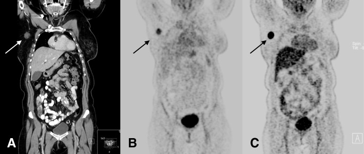
Fig. 2.
67-year-old patient with a nodal recurrence 22 months after treatment of a primary breast cancer. Coronal CT reconstruction shows a contrast enhancing lymph node metastasis with a diameter of 2.1 cm in the right axillary region (a). The lesion is visually detectable on 68Ga-Pentixafor PET with a corresponding SUVmax of 4.0 (b). On 18F-FDG PET/CT, the lesion shows a significantly higher tracer uptake (SUVmax of 24.4) (c)

