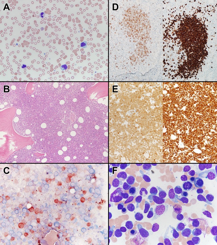
Fig. 1.
a Peripheral blood smear of the patient with a diagnosis of chronic myelomonocytic leukemia, demonstrating atypical dysplastic granulocytes, atypical monocytes, and a blast. Wright Giemsa stain, ×400 magnification. b Bone marrow core biopsy of the patient with chronic myelomonocytic leukemia, demonstrating a hypercellular marrow (90%), with dysplastic megakaryocytes. Hematoxylin and eosin, ×100 magnification. c Butyrate esterase (brown) and chloroacetate esterase (blue) cytochemical dual stain, demonstrating increased butyrate esterase monocytes and dual esterase-positive bone marrow monocytes, suggestive of monocytic dysplasia (×400 magnification). d (Left) Bone marrow core biopsy, TCL1 immunohistochemistry, demonstrating TCL1-positive plasmacytoid dendritic cell nodules (×200 magnification). (Right) Bone marrow core biopsy, CD123 immunohistochemistry, demonstrating CD123-positive plasmacytoid dendritic cell nodules (×200 magnification). e (Left) Bone marrow biopsy, TCL1 immunohistochemistry, demonstrating diffusely TCL1-positive blasts, suggestive of a blastic plasmacytoid dendritic cell neoplasm (×100 magnification). (Right) Bone marrow core biopsy, CD123 immunohistochemistry, demonstrating diffusely positive CD123 blasts, suggestive of a blastic plasmacytoid dendritic cell neoplasm (×100 magnification). f Bone marrow aspirate obtained at the time of disease transformation demonstrating hand mirror-shaped blastic cells, characteristic for blastic plasmacytoid dendritic cell neoplasms. Wright Giemsa, ×1000 magnification

