
Figure 3.
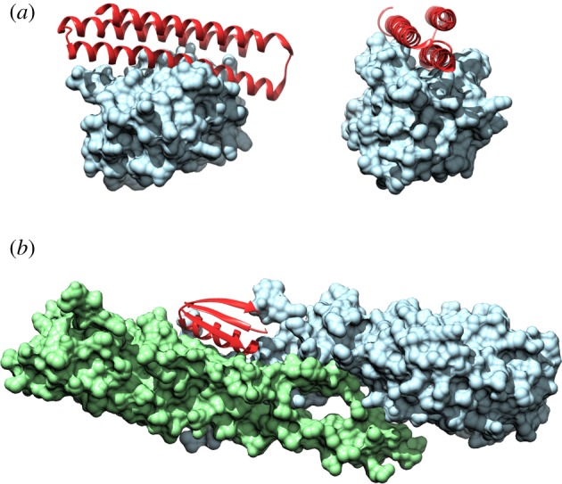
Crystal structures of de novo inhibitors binding to their targets. Crystal structures of inhibitor complexes. (a) Inhibitor peptide αMCL1 (red) binds the human BCL2 homologue, Mcl-1 (blue), with picomolar affinity [60]. PDB: 5JSB. (b) Inhibitor peptide HB1.6928.2.3 (red), which can bind influenza haemagglutinin [59]. PDB: 5VLI.