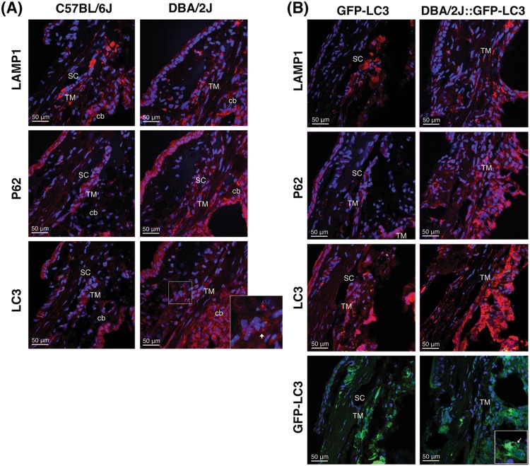
Fig. 3. Immunofluorescence staining of autophagy lysosomal markers in the iridocorneal angle region of C57BL/6J and DBA/2J mice.
Representative immunofluorescence staining of LAMP1, p62, and LC3 in the angle region of non-GFP (a) and GFP-LC3 transgenic mice (b). Red: specific antibody staining; blue: DAPI; green: GFP-LC3 fluorescence. SC Schlemm’s canal, TM trabecular meshwork, cb ciliary body. Arrows indicate the presence of LC3 puncta

