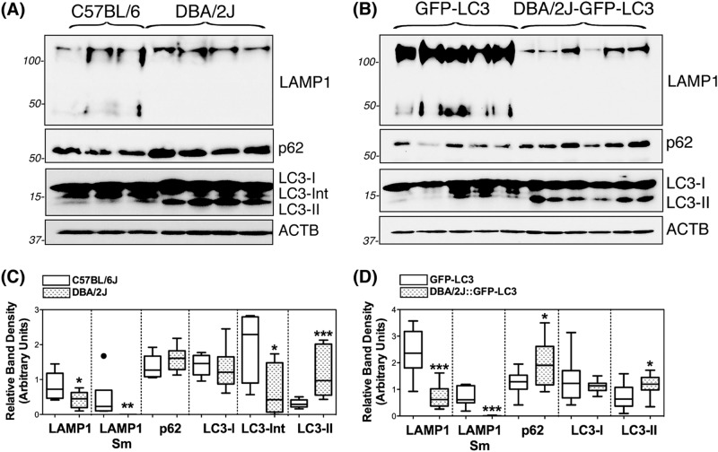
Fig. 4. Western-blot analysis of autophagy lysosomal markers in dissected iridocorneal angle regions of C57BL/6J and DBA/2J mice.
Protein expression levels of LC3, p62, and LAMP1 in whole-tissue lysates (5–10 μg) of dissected iridocorneal angle region from non-GFP (a) and GFP-LC3 transgenic mice (b) analyzed by western blot; c and d represent the normalized relative protein levels calculated from densitometric analysis of the blots. Data are the means ± SD; *p < 0.05, **p < 0.01, ***p < 0.001, Tukey test; nC57BL/6J = 6, nDBA/2J = 9, nGFP-LC3 = 10, nDBA/2J::GFP-LC3 = 10. Sm: small band, non-glycosylated form

