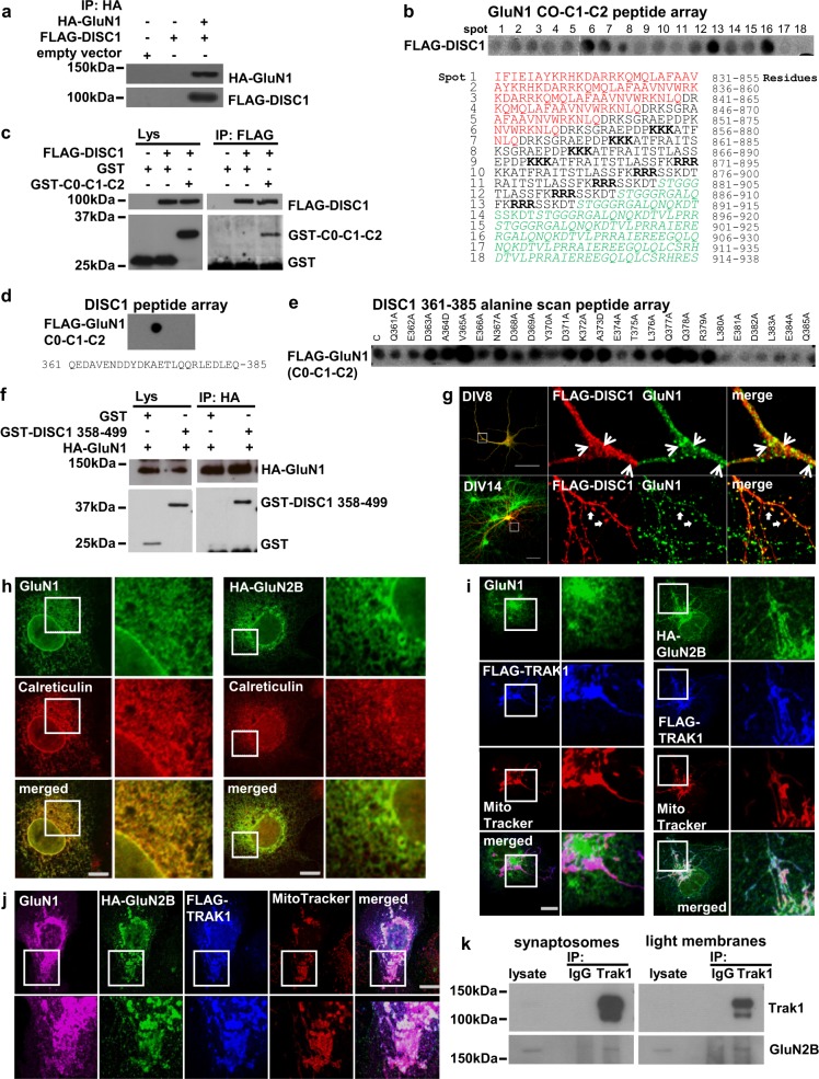
Fig. 3. DISC1 interacts with GluN1 and TRAK1 associates with GluN2B.
a HA–GluN1 co-immunoprecipitates FLAG-DISC1 from transfected COS7 cells. b GluN1 cytoplasmic tail (C0-C1-C2) peptide array hybridised with Flag-DISC1. Each spot represents a single peptide. Red, black and green texts mark the C0, C1 and C2 cassettes, respectively. ER retention signals are in bold. c FLAG-DISC1 co-immunoprecipitates GST-tagged C0-C1-C2. d DISC1 peptide array probed with FLAG-C0-C1-C2. e DISC1 361–385 GluN1 binding region alanine scan probed with FLAG-C0-C1-C2. C, unmutated peptide. f HA–GluN1 co-immunoprecipitates GST-DISC1 amino acids 358–499 from transfected COS7 cells. g Co-localisation of FLAG-DISC1 and endogenous GluN1 in cultured DIV8 and DIV14 hippocampal neurons. Arrowheads and arrows indicate example sites of co-localisation. h COS7 cells transfected with GluN1 or HA–GluN2B expression constructs were labelled with antibodies specific for GluN1 or HA, plus the ER marker Calreticulin. i COS7 cells co-transfected with FLAG-TRAK1 plus GluN1 (left) or HA–GluN2B (right) expression constructs were labelled using antibodies specific for GluN1 or HA, plus anti-FLAG and the mitochondrial dye Mitotracker CMXRos. j COS7 cells triple transfected with FLAG-TRAK1, GluN1 plus HA–GluN2B expression constructs were labelled using antibodies specific for GluN1, HA and FLAG, plus Mitotracker CMXRos. COS7 cells were used because they are ideal for exogenous protein expression due to their high transfection efficiency, large size and low profile, which facilitate co-immunoprecipitation and co-localisation studies to complement endogenous protein studies in neurons. k Trak1 immunoprecipitates GluN2B from adult mouse brain synaptosome and light membrane fractions. Scale bars, 50 μm in G, otherwise 20 μm; white boxes indicate enlarged areas

