Figure 4.

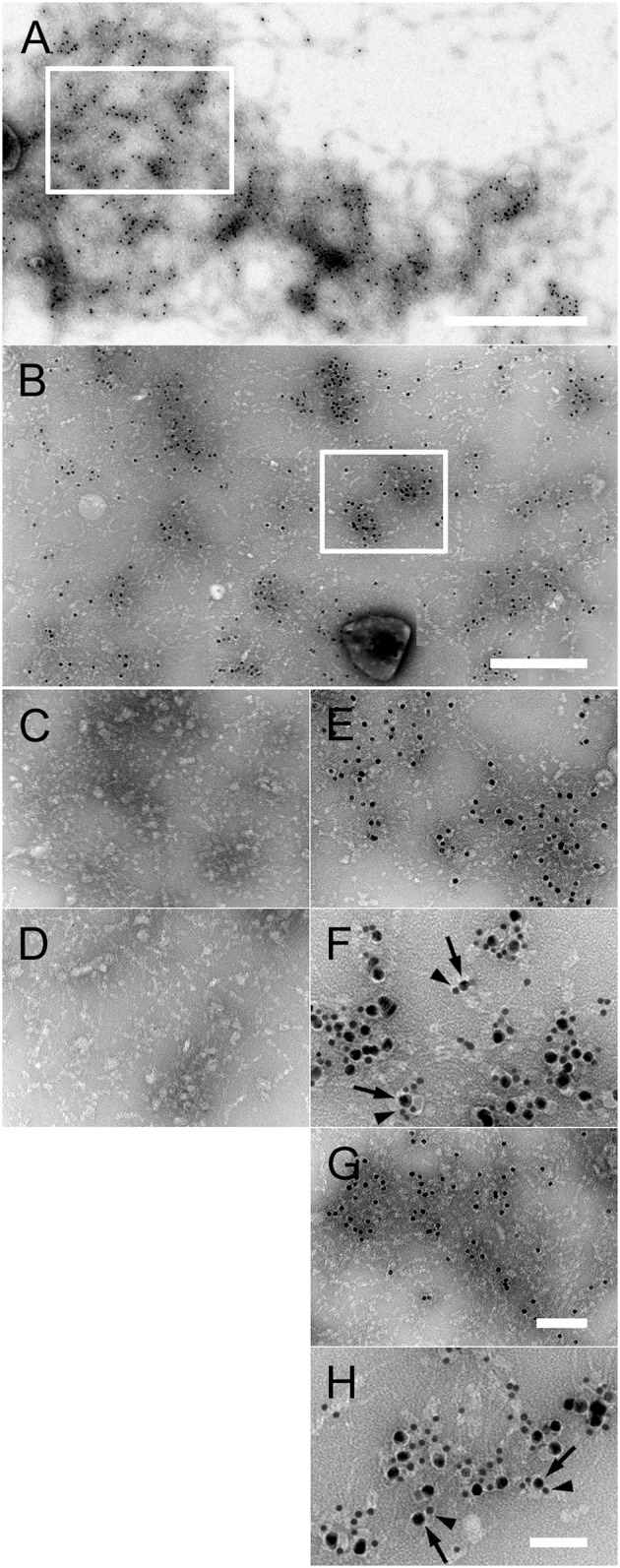
Interaction of purified PE and Hap with collagen VI networks in vitro. (A) Overview of extended native collagen VI networks from bovine cornea after incubation with gold-labeled PE. The 10 nm gold-PE conjugates are barely visible as black dots. The scale bar represents 500 nm. (B) Enlargement of an area corresponding to the frame in (A) exhibiting extensive binding of 10 nm gold-labeled PE to the collagen VI network. Scale bar, 250 nm. (C–H) Enlargements of collagen VI networks incubated with the different gold-labeled NTHi surface proteins as indicated by the frame in (B). (C) Collagen VI alone, (D) Hia, (E) PE, (F) colocalisation of PE (10 nm) with collagen VI VWA domains (5 nm), (G) Hap, (H) colocalisation of Hap (10 nm) with collagen VI VWA domains (5 nm). The scale bars represent 100 nm (C–E,G) and 50 nm (F,H).
