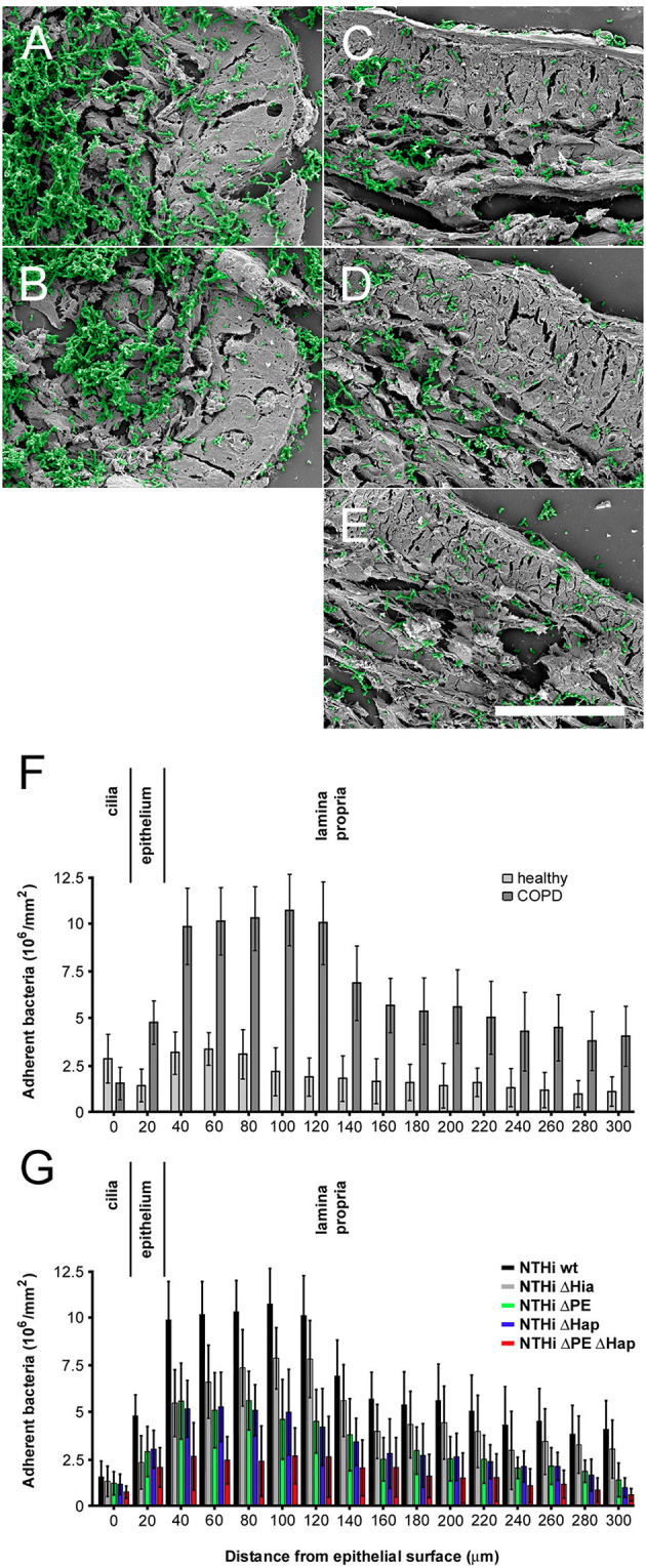
Figure 5.

Colonization of NTHi in COPD airways ex vivo. Scanning electron micrographs of paraffin sections of airway biopsies from COPD patients inoculated with NTHi wild type (A), Δhia (B), Δhpe (C), Δhap (D), and ΔhpeΔhap (E). In COPD, considerable numbers of wild type and Δhia bacteria are observed bound to the airway submucosa (A,B) as compared to bacteria lacking PE (C), Hap (D), or both adhesins (E). Bacteria are highlighted in green pseudocolour. The scale bar represents 50 μm. (F,G) Quantitative evaluation of adherent bacteria as indicated.
