Figure 6.

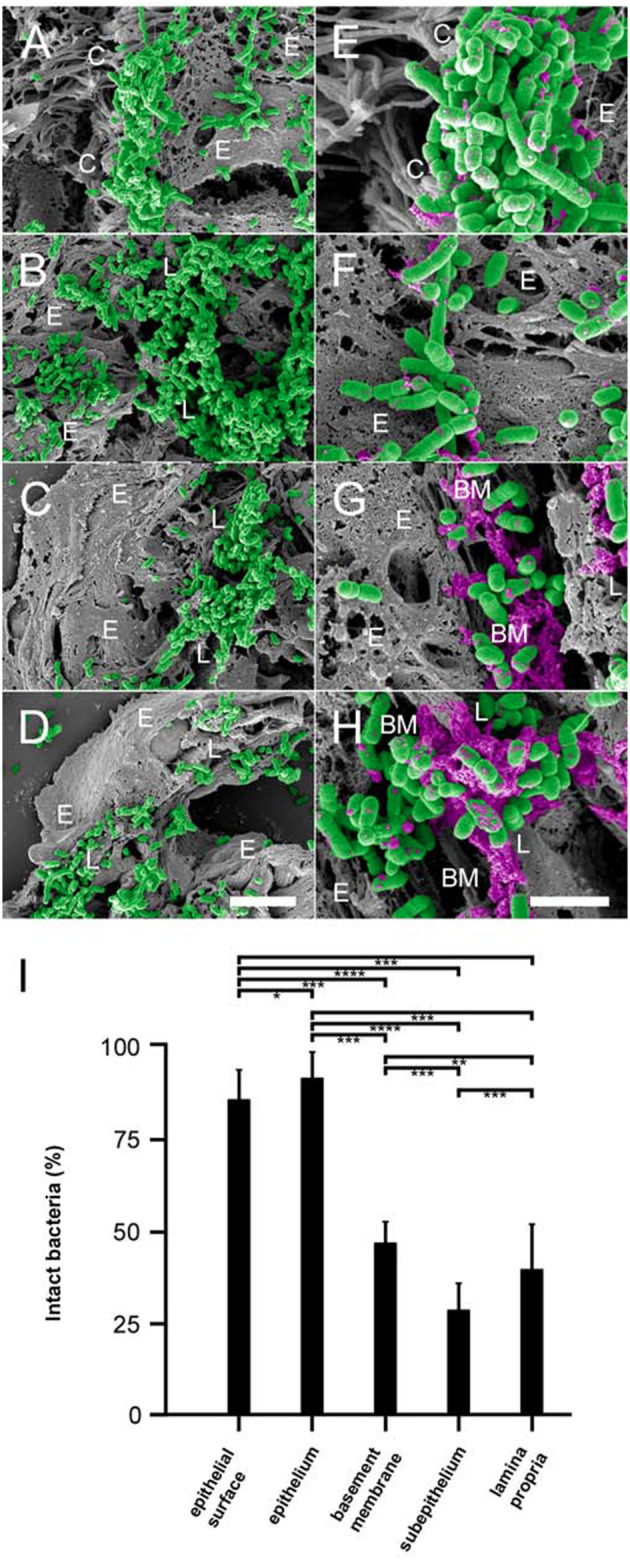
Colonization of pulmonary compartments by NTHi and antimicrobial activity of the lamina propria in vivo. Paraffin sections of murine COPD lungs inoculated with NTHi by intratracheal challenge. Scanning electron microscopy of large airways reveals extensive binding of bacteria to the epithelial surface (A) and the lamina propria (B). Similar observations were made for small airways (C) and alveoli (D), except that bacteria colonize the subepithelial lamina propria and bind only sparsely to the epithelial surface. (E–H) Higher magnification of the same areas reveals killing of NTHi bacteria upon exposure to the epithelial surface (E), the epithelial cell layer (F), the subepithelial basement membrane (G), and the lamina propria (H). Bacteria are highlighted in green and cytoplasmic exudates in purple pseudocolour. Cellular and tissue structures are indicated as follows; C, epithelial cilia; E, epithelial cell; L, lamina propria; BM, basement membrane. The scale bars represent 5 μm (A–D) and 2 μm (E–H). (I) Quantitative evaluation of bacterial killing as indicated.
