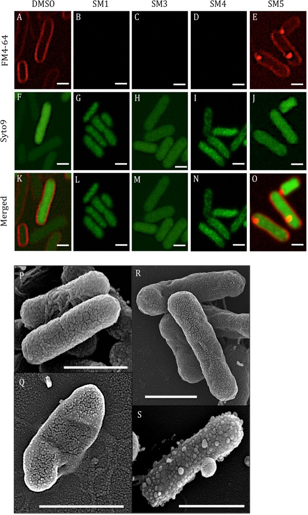
Figure 3.
Confocal and scanning electron microscopy (SEM) analyses of Salmonella enterica subsp. enterica serotype Typhimurium after challenge with five times the minimal bactericidal concentraiton of small molecules (5X MBC of SMs) for 3 hrs. (A–O) Confocal microscopy: (A–E) S. Typhimurium cell membrane stained with FM4-64; (F–J) S. Typhimurium nucleic acids stained with SYTO9; (K–O) Merged pictures of the FM4-64 and SYTO9 staining. (P–S) SEM: (P) 1% dimethyl sulfoxide (DMSO) treated Salmonella; (Q) SM1 treated Salmonella; (R) SM4 treated Salmonella; (S) SM5 treated Salmonella; Bar: 1 μm.

