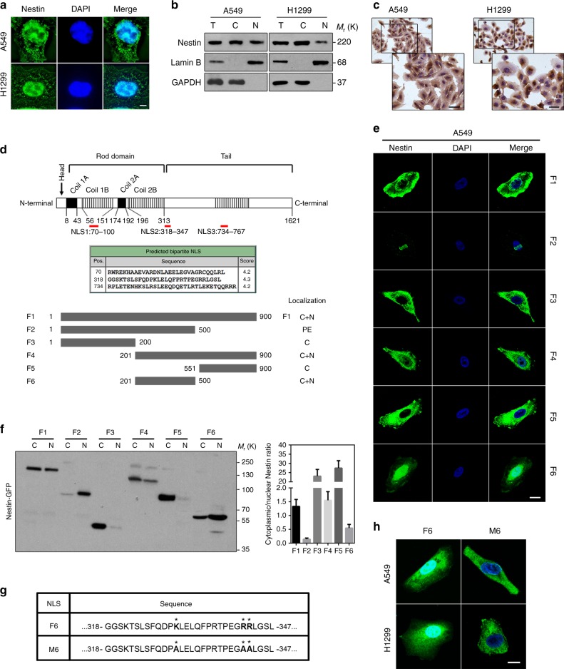
Fig. 3.
Nestin contains a nuclear localization signal (NLS). a Confocal images of Nestin immunostaining (green) in A549 and H1299 cells. Scale bars, 5 μm. b Immunoblotting for subcellular fractions of Nestin in A549 and H1299 cells (T total cell lysates, C cytoplasmic cell lysates, N nuclear cell lysates). c Immunohistochemical staining of Nestin in A549 and H1299 cells. Scale bars, 30 μm. d Upper: three putative NLSs identified by bioinformatic analysis. Lower: schematics of the Nestin constructs used for the localization study. Each fusion protein was expressed with a GFP tag (left), and its localization was assessed (right; C, predominantly cytoplasmic; C + N, in both compartments; PE, at the periphery of outer nuclear envelope). e Confocal images of GFP-Nestin fusion protein in A549 cells. Nuclei were marked by DAPI (blue). Scale bars, 20 μm. f Immunoblotting analysis of GFP-Nestin distribution in A549 cells (left; C cytoplasmic cell lysates, N nuclear cell lysates) and the corresponding cytoplasm/nucleus intensity ratio (CN ratio) of GFP-tagged proteins (right). g, h Schematics of the GFP-tagged Nestin constructs showing the sequences (g) and positions (h) of a potential NLS and the targeted aa substitutions (asterisk) used to disable it. Scale bars, 10 μm

