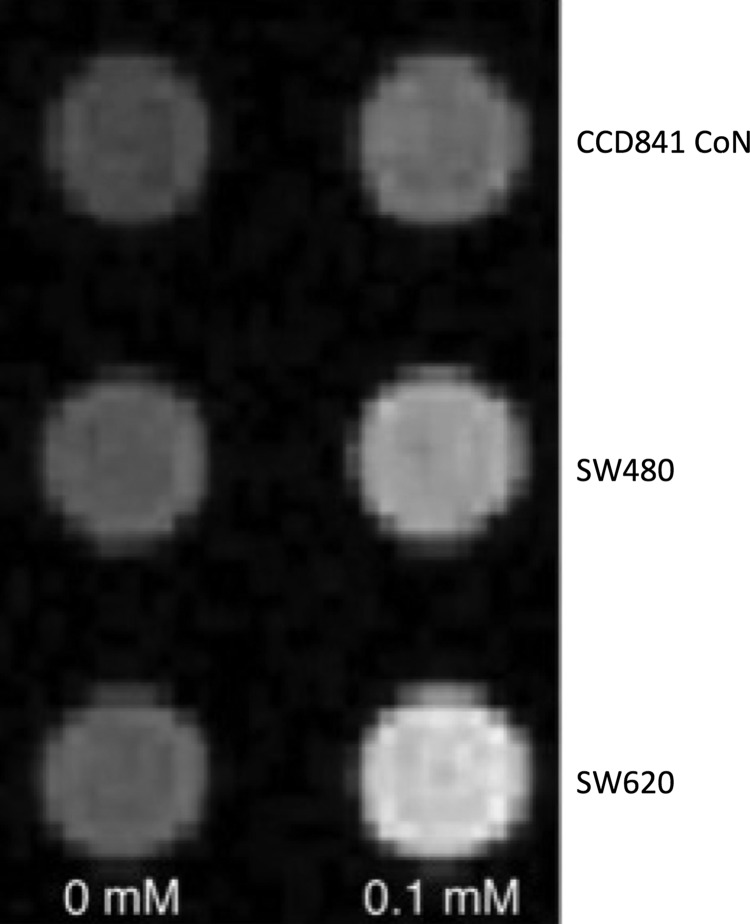
Figure 1.
In vitro manganese-enhanced magnetic resonance imaging (MEMRI) of SW620, SW480, and normal cell lines. T1-weighted image (T1WI) of cell pellets from cells exposed to a 0- or 0.1 mM MnCl2 incubated for 60 min revealed both cancer cells and normal cell were enhanced by various degrees; however, cancer cells displayed a more marked level of enhancement versus the normal cellular counterpart.

