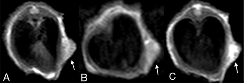
Figure 2.
2D turbo spin echo (2D TSE) T1WI of an SW620 xenograft in the 5-mm tumor group (arrow). In the unenhanced scan, the tumor manifests as a mildly high signal intensity relative to muscle (A). In the conventional gadolinium (Gd)-enhanced scan, the tumor enhances slightly and inhomogeneously (B). In the Mn-enhanced scan, more prominent and a larger extent of enhancement is readily noted (C).

