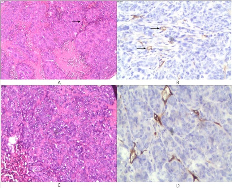
Figure 5.
Histopathological photomicrograph of an SW480 tumor in the 10-mm-size tumor group (A and B). Hematoxylin and eosin (H&E) staining 40× (A). Packed tumor cells with few interstitia (black arrow) and discrete coagulative necrosis (white arrow) are shown. CD34 immunohistochemical staining 200× (B). Sparse microvessel staining (arrows) are shown. Histopathological photomicrograph of an SW620 tumor in the 5-mm-size tumor group (C and D). H&E staining 100× (C). Few interstitia (white arrow) within diffuse tumor cells are shown. CD34 immunohistochemical staining 200× (D). Arrows point to sparse microvessels.

