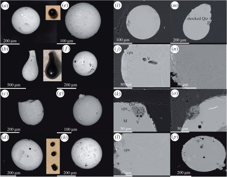
Figure 2.
Electron backscatter (15 kV) images of some representative P-E spherules (microtektites and microkrystites) from Hole 1051B, Wilson Lake B, and Millville cores (modified from Schaller et al. [18]). Insets are light micrographs. (i–p) show P-E boundary spherule cross sections mounted in epoxy (modified from Schaller et al. [18]). Note the classic dendritic quench textures. Shocked quartz grain in (m) is shown in detail in figure 6. See Schaller et al. [18] for details. (Online version in colour.)

