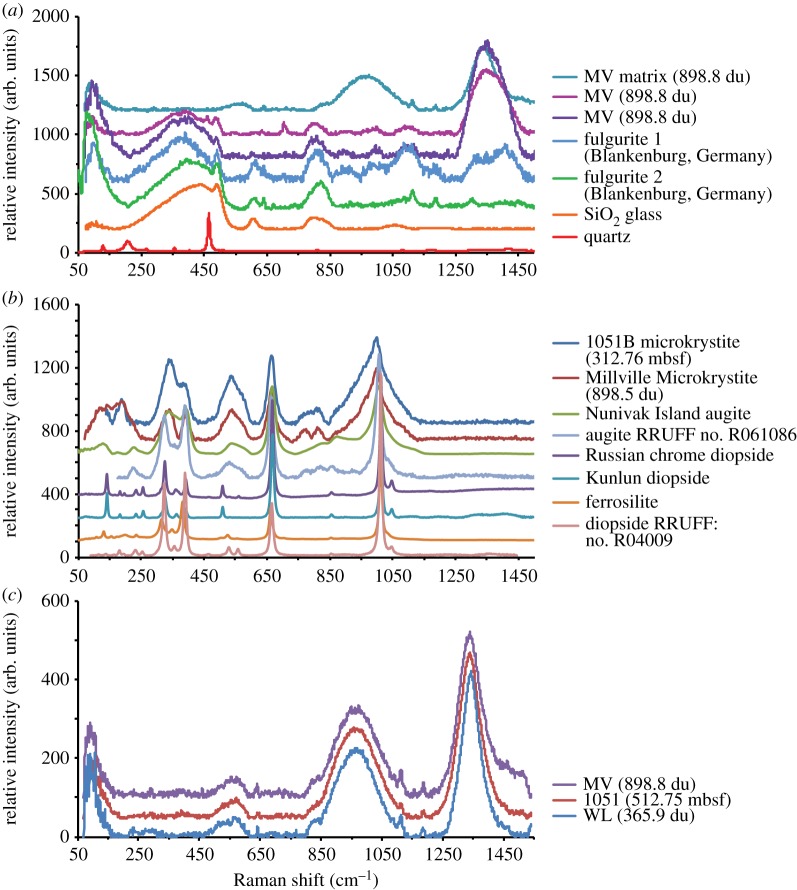
Figure 5.
(a) Background corrected Raman spectra of representative lechatelierite inclusions found in PE spherules compared with the spherule matrix, lechatelierite from fulgurite, SiO2 glass, and quartz (modified from Schaller et al. [18]). (b) Raman spectra of representative clinopyroxene crystallites compared with augite, diopside and ferrosilite standards [18]. (c) Micro-laser Raman spectra of representative microtektite matrices. Spectra are collected using a Bruker 532 nm green laser system at the Rensselaer Polytechnic Institute, Troy, NY, and are background corrected [18].

