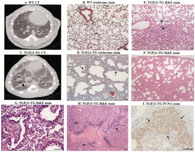
FIGURE 3.
Pulmonary fibrosis in transgenic (TG) mice overexpressing human transforming growth factor (TGF)-β1. CT and trichrome stain of lung tissues from WT mice (A,B) and TGF-β1 mice (C,D, arrows). H&E stain showed areas of fibrotic changes alternate with areas with less affected parenchyma (E, arrows), honeycombing-like cysts with disruption of lung parenchyma (F), and fibroblastic foci-like structures (G, arrow), and multiples areas with adenocarcinoma (H, arrows). There is increased staining of proliferating cell nuclear antigen (PCNA) in cancerous areas (I, arrows). Scale bars indicate 50 μm.

