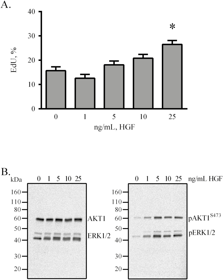
Figure 2.
Hepatocyte growth factor stimulates proliferation and phosphorylation of AKT1 and ERK1/2. Semi-confluent eqSC (n = 5) were treated with HGF for 48 h with a 2-h pulse of EdU prior to fixation (A). Cells were treated with the indicated concentration of HGF for 20 min, lysed and analyzed by Western blot for total and phosphorylated forms of AKT1 and ERK1/2 (B). EdU (+) and total nuclei were enumerated. Western blot exposure time was 2 mins. Percent EdU = EdU (+)/Hoechst 33342 (+) × 100. Means and SEMs shown. *Significance between control and treatment at P < 0.05. AKT1 = AKT serine/threonine kinase 1; eqSC = equine satellite cell.

