Figure 5.
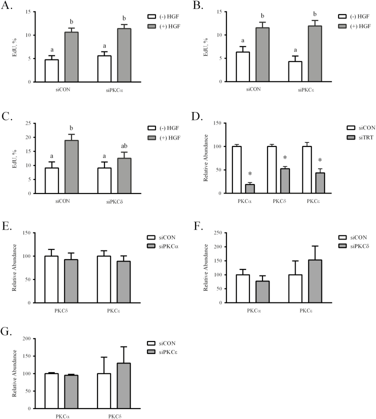
Hepatocyte growth factor signaling through PKCδ stimulates eqSC proliferation. Semi-confluent eqSC (n = 5) were transfected with scrambled (siCON) or siRNA specific for PKC isoforms. After 24 h, the cells were treated with HGF for 24 h with a 2 h EdU pulse prior to fixation. Total and EdU (+) nuclei were enumerated. Equine SC treated with siPKCα (A) or siPKCε (B) did not prevent HGF stimulated proliferation. Cells expressing siPKCδ did not respond to HGF (C). Means with different letters are significant at P < 0.05. Total RNA from siCON- and siPKC-treated cells was analyzed by qPCR for specific knockdown (D) and off-target (E–G) reduction of PKCα, PKCδ, and PKCε mRNA. Relative expression was calculated by 2−ΔΔCt method. *Significance at P < 0.05 within treatment. eqSC = equine satellite cell; qPCR = quantitative PCR; siRNA = small interfering RNA.

