
Figure 4. hESCs cultured in m5i/LAF are hypomethylated and have two active X chromosomes.
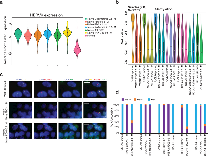
(a) Violin plot representation of HERVK expression (n=64) in hESCs expanded in the indicated culture conditions (P10). For primed, naïve PD03 1μM and naïve PD03 0.5 μM n=5 independent hESC lines (UCLA1, 4, 5, 9 and WIBR3), for naïve Cobimetanib 0.5 μM, Refametinib, TAK-733 and t2iLGöY n=1 (UCLA4). (b) Global methylation analysis of primed and naive hESCs by RRBS analysis using violin plot representation. (c) Representative RNA FISH images for primed and naïve hESCs at P16, detecting the indicated transcripts. (scale bar: 10 μm). (d) Quantification of XIST RNA FISH patterns.