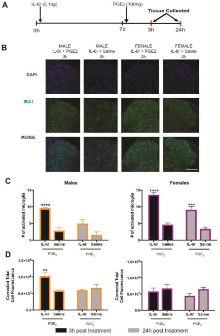
Figure 2. Hyperalgesic priming revealed with PGE2 injection after IL-6r leads to increased spinal microglia at 3 hr after injection.
A. Animals received an intraplantar injection of IL-6 and then 7 d later received an intraplantar injection of PGE2. Spinal cords were dissected at 3 or 24h post (n = 4–6 mice per group) B. After PGE2 injection spinal cords were stained for IBA1 and DAPI. Scale bar is 200 μm. C. 3 hr following PGE2 injection in primed mice, levels of activated microglia were elevated in both males and females. D. Total cell fluorescence was only elevated in male mice at 3 hr post PGE2 injection. Data are displayed as mean ± SEM. Differences between groups were measured using a two-Way ANOVA with Bonferroni’s post hoc test, **p < 0.005, ***p < 0.0005, ****p < 0.0001.

