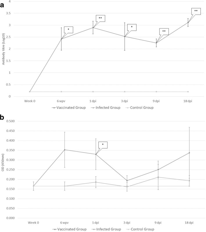
Fig. 1.
Plasma levels of specific rFhCL1 IgG1 (a) and IgG2 (b). Each point represents mean values of antibody titre - log10 - (IgG1) and of optical density (IgG2) measured at 450 nm. Bars at each point represents tandard error. Immunisation with rFhCL1 developed a significant rise in the level of IgG1 isotype; dynamics of IgG1 in uninfected and infected sheep (Groups 1 and 2) shows a similar pattern, hence it is overlapped in the figure. Significant IgG2 production was detected only at 1 day post-infection (dpi) during the trial

