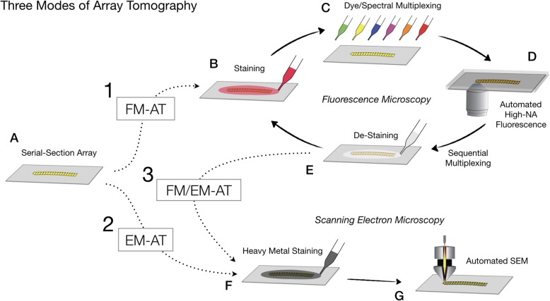
Fig. 2.
Alternative fluorescence (FM-AT), electron (EM-AT) and combined (FM/EM-AT) modes of array tomography. An array of serial ultrathin sections (a) (e.g., a single-ribbon coverslip produced as in Fig. 1) may be stained and imaged for multiplex fluorescence microscopy (dotted arrow 1), scanning EM (dotted arrow 2), or both (dotted arrows 1 and 3). (b–e) Schematizes possible combinations of spectral and sequential fluorescence multiplexing modes. (f, g) Schematizes (optional) array staining and image acquisition for electron microscopy

