Fig. S6.
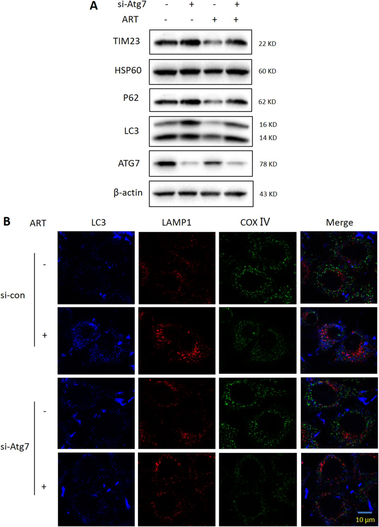
Suppl Fig. 6. ATG7 depletion suppresses mitophagy. (A) Western blot analysis of mitophagy levels in ART-treated HeLa cells after Atg7 knockdown. The cells were first transiently transfected with a nonspecific siRNA or the Atg7-specific siRNA and were subsequently treated with 10 μM ART for 12 h. Cell lysates were then prepared and subjected to western blotting analysis for mitochondrial proteins TIM23, HSP60, LC3, P62 and ATG7. β-actin was used as the loading control. (B) Fluorescence microscopy of mitophagy activity in ART-treated HeLa cells after Atg7 knockdown. The cells were prepared for immunostaining with LC3 (Pacific Blue), LAMP1 (Alexa Fluor 594, red) and COX Ⅳ (Alex Fluo 488, green) and then examined under confocal microscopy. Scale bar: 10 µm.

