Fig. S7.
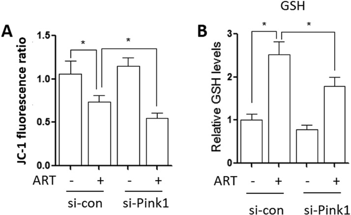
Suppl Fig. 7. Inhibition of mitophagy alters mitochondrial function. (A) Mitochondria membrane potential of ART-treated HeLa cells after PINK1 knockdown. The cells were first transiently transfected with the PINK1-specific siRNA and then probed with JC-1 (10 μg/ml) to assess mitochondrial membrane potential. Flow cytometry was performed to determine cell fluorescence. * P < 0.05 (B) Glutathione levels in ART-treated HeLa cells after PINK1 knockdown. The cells were first treated as per (A) and incubated with ThiolTracker™ Violet dye (20 µM) at 37 °C for 30 min. Cell fluorescence was measured by flow cytometry at 405 nm excitation. * P < 0.05.

