
Figure 4.
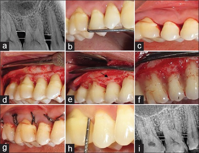
Group C (OFD + IMP + PRFM) (a) radiovisiography baseline-linear radiographic interpretation with AutoCAD computer software (parallel lines showed the base of the defect and crest of alveolar bone); (b) probing pocket depth; (c) incisions placed; (d) debridement done along with intra marrow penetration; (e) direct interrupted suture placed; (f) probing pocket depth at 9 month; (g) radiovisiography 9 months-linear radiographic interpretation with AutoCAD computer software (parallel lines showed the base of the defect and crest of alveolar bone). OFD + IMP + PRFM – Open flap debridement with intramarrow penetration and platelet-rich fibrin matrix gel