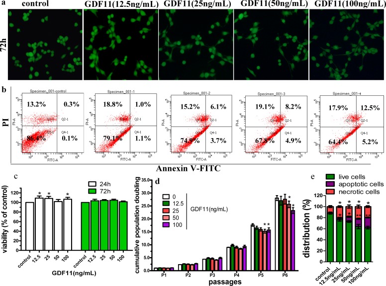
Figure 1. Effect of GDF11 on C17.2 cells.
(A) The representative images of live and dead cell staining. C17.2 cells were cultured with indicated concentrations of GDF11. Images were obtained at 200× magnification by inverted fluorescence microscope. The live cells were stained with calcein AM in green, and the dead cells were stained with EthD-1 in red. (B) GDF11 induced apoptosis in C17.2 neural stem cells. C17.2 cells were treated with vehicle or GDF11 (12.5, 25, 50 and 100 ng/mL) for 48 h and cell distribution was analysed using Annexin V-FITC and PI dual staining. The FITC and PI fluorescence was measured by flow cytometer with FL-1 and FL-2 filters, respectively. Lower left quadrant–live cells (Annexin V−/PI−), lower right quadrant–early/primary apoptotic cells (Annexin V+/PI−), upper right quadrant–late/secondary apoptotic cells (Annexin V+/PI +) and upper left quadrant–necrotic cells (Annexin V−/PI+). (C) The viability of C17.2 cells after 24 h or 72 h of cultivation with various concentrations of GDF11 or vehicle was measured using CCK-8 method. N = 3, p < 0.05. (D) Cumulative population doubling levels of C17.2 cells supplemented with different GDF11 concentrations for a total period of 6 passages. N = 4, *p < 0.05 compared with control. (E) Quantitative analyses of the GDF11 effect on apoptosis. N = 3, *p < 0.05 versus with vehicle control and **p < 0.01 versus with vehicle control.

