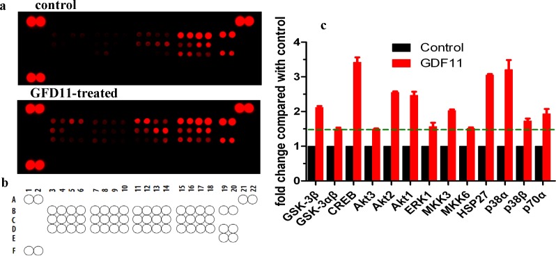
Figure 5. Alterations of MAPK pathway-related proteins in GDF11-treated C17.2 cells.
(A) Phosphoproteome profiling of C17.2 cells in response to GDF11 stimulation. Total cell lysates from C17.2 cells with25 ng/mL GDF11- or vehicle-treated were incubated on membranes of the phospho-proteomics platforms (human Phospho-MAPK, 23 different MAPKs and other serine/threonine kinases), as described in “Materials and Methods”. (B) Human Phospho-MAPK array coordinates. (C) The graph shows the relative fold change of proteins with significant difference upon GDF11 treatment, setting 1 for control. Protein levels with higher than ±1.5 folds indicated by dotted lines are considered as the differentially expressed proteins.

