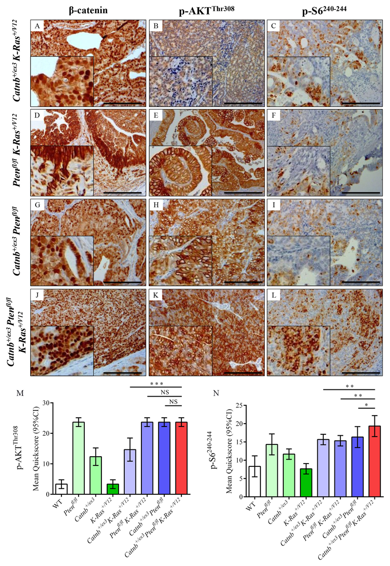
Figure 5. Immunohistochemistry for β-catenin, p-AKTThr308 and p-S6240-244 in double and triple allele mouse models.
(A-L) Immunostaining for β-catenin (A, D, G, J), p-AKTThr308 (B, E, H, K), and p-S6240-244 (C, F, I, L) on Pb-Cre4+ Catnb+/ex3 K-Ras+/V12 (A-C), Pb-Cre4+ Ptenfl/fl K-Ras+/V12 (D-F), Pb-Cre4+ Catnb+/ex3Ptenfl/fl (G-I) and Pb-Cre4+ Catnb+/ex3Ptenfl/flK-Ras+/V12 (J-L) cohorts of mice (n=4) at the endpoint of the experiment (500 days or when sick. Bars = 200 μm. Insets magnified two times. (M, N) Quickscore quantification of p-AKTThr308 and p-S6240-244 for all genotypes. For Quickscore quantification of β-catenin membrane-specific (M) and nuclear staining (N), see Fig.4M,N. For clarity, significance of Quickscore staining in triple mutant vs double mutant cohorts only is presented on the graphs (NS, not significant;* P<0.05; ** P<0.01; *** P<0.001; unpaired two-tailed t-test, n=4); full statistical comparisons of all genotypes are given in Supplementary Table 1. Catnb+/ex3K-Ras+/V12 tumours demonstrated predominantly nuclear staining of β-catenin, with weak focal staining for p-AKTThr308 and p-S6240-244 (A-C). Ptenfl/fl K-Ras+/V12 tumours had strong membrane staining for β-catenin, strong diffuse staining for p-AKTThr308 and focal positive staining for p-S6240-244 (D-F). Catnb+/ex3Ptenfl/fl tumours demonstrated both nuclear and membrane-specific β-catenin staining, diffuse staining for p-AKTThr308 and focal positive staining for p-S6240-244 (G-I). Catnb+/ex3Ptenfl/flK-Ras+/V12 tumours stained avidly for nuclear β-catenin with diffuse strong staining for both p-AKTThr308 and p-S6240-244 (J-L), the latter being significantly higher than in all other cohorts.

