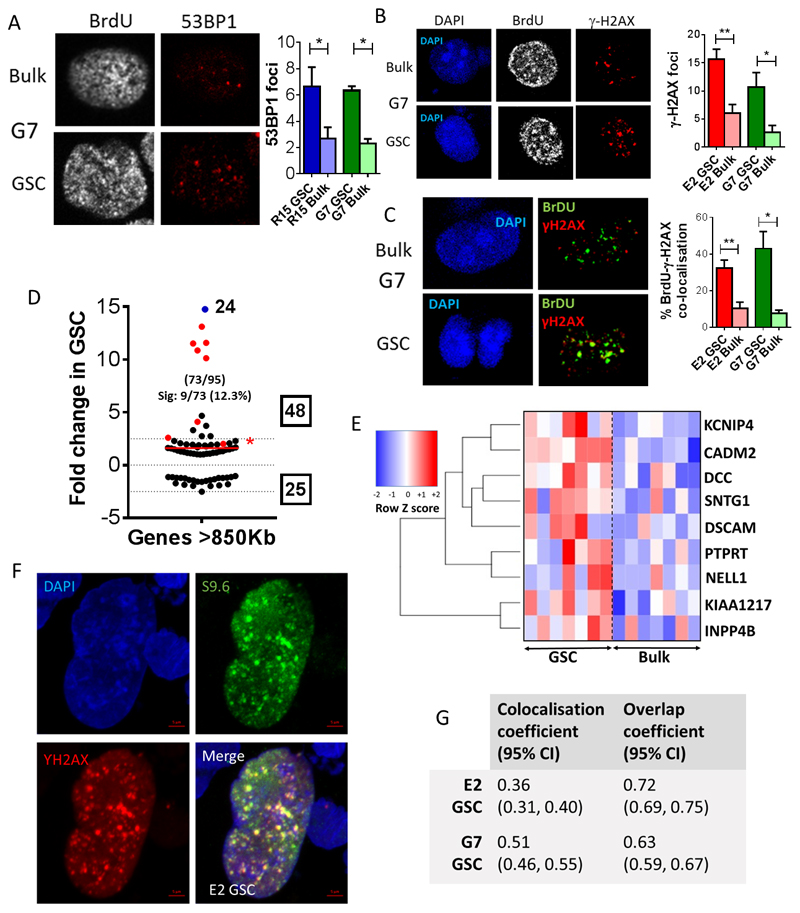
Figure 3. GSCs demonstrate increased numbers of γ-H2AX foci, which co-localize with replication factories and RNA: DNA hybrids.
A-B Representative immunofluorescence images of G7 GSC and tumor bulk cells showing (A) 53BP1 and (B) γ-H2AX foci in BrdU positive cells under basal conditions, with quantification of (A) 53BP1 and (B) γ-H2AX foci per S-phase nucleus in G7 and E2 GSC and tumor bulk cells (mean +/-SEM, n = 3, *p<0.05, **p<0.01). C Representative images demonstrating co-localization of γ-H2AX foci with BrdU replication factories (BrdU foci) in G7 GSC and tumor bulk cells. Percentages of BrdU positive replication factories co-localizing with γ-H2AX foci are quantified in E2 and G7 GSC and tumor bulk cells (mean +/-SEM, n=3, *p<0.05, **p<0.01, unpaired t-test). D Mean fold change in the expression of genes across 7 GSC cultures compared to the paired tumor bulk cells associated with genes >850kb in length. Numbers of genes identified from the RNA sequencing data and total numbers of genes in the published gene dataset is shown in brackets and total numbers of up-and down regulated genes are indicated in boxes. The numbers and percentages of significantly altered (‘Sig’) genes in each dataset are shown and these genes are highlighted in red. Gene shown in blue was up regulated 24-fold. Mean fold changes across all genes are shown by red lines. Genes >850bp in length are significantly up regulated in GSC compared to paired tumor bulk population across 7 GBM cell lines (one sample t-test, *p<0.05, NS=non-significant). E Heatmap illustrating fold changes in expression of the 9 significantly up regulated genes >850bp across 7 paired cell lines. F Representative image of immunofluorescent staining for RNA: DNA hybrids using S9.6 antibody and γ-H2AX in E2 GSC. G Table of colocalization and overlap coefficients (95% CIs) for γ-H2AX versus S9.6 immunofluorescence in E2 and G7 GSC.

