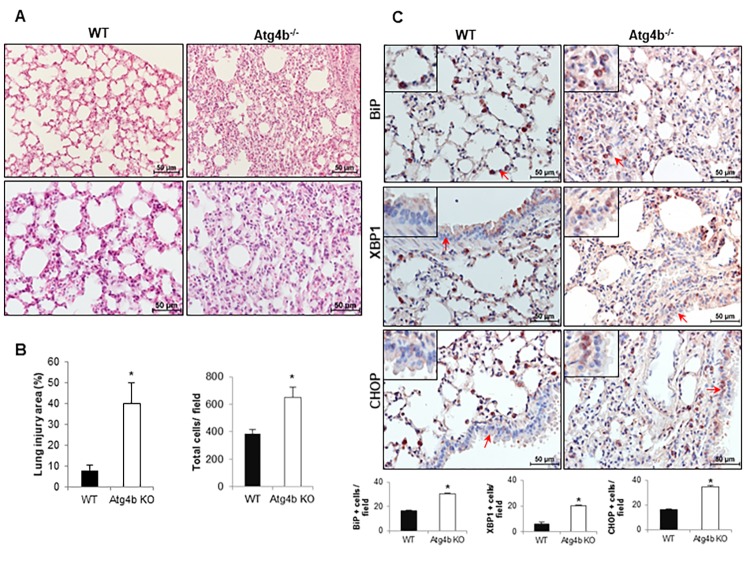
Figure 4.
Exacerbated inflammatory and ER stress response in lungs from Atg4b null mice. (A) Representative light microscopy images of H&E stained lung tissue sections from WT and Atg4b null mice after 3 days post-tunicamycin treatment. (B) Lung injury area and total cell number in the lung fields by quantitative image analysis. (C) Representative light microscopy images of BiP, XBP1 and CHOP positive stained lung tissue sections from WT and Atg4b null tunicamycin-treated mice at 3 days (top panels). All insets show 2X larger magnification. Total number of positive stained cells per high power field (40X) in lung tissue sections by quantitative image analysis (bottom panels). Results shown represent mean ± SD. Statistical significance was determined by Student’s t test (*p < 0.05).

