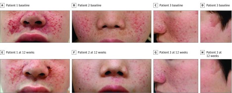
Figure 3. Representative Photographs From Patients in the Sirolimus Group.
A-C, Angiofibromas on the cheek at baseline in patient 1, a teenage boy; patient 2, a young boy; and patient 3, a man in his 20s. D-F, After 12-week treatment with sirolimus gel, 0.2%, the angiofibromas were rated improved, improved, and markedly improved, respectively, showing reduced size and faded color. G and H, A cephalic plaque on the temple of patient 3 at baseline appeared flattened and was rated improved after 12-week treatment with sirolimus gel, 0.2%.

