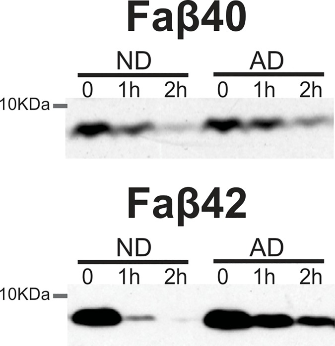
Fig 1. Representative western blot showing faster Aβ degradation in the liver of non-demented controls subjects when compared to the AD group.
Lyophilized fluorescein-labeled Aβ40 (A) or Aβ42 (B) peptides were added to liver homogenates to quantify its degradation. Sucrose lysis buffer containing complete protease inhibitor cocktail was added to stop the reaction and Western blot performed to visualized Aβ degradation (molecular weight ~7kDA).

