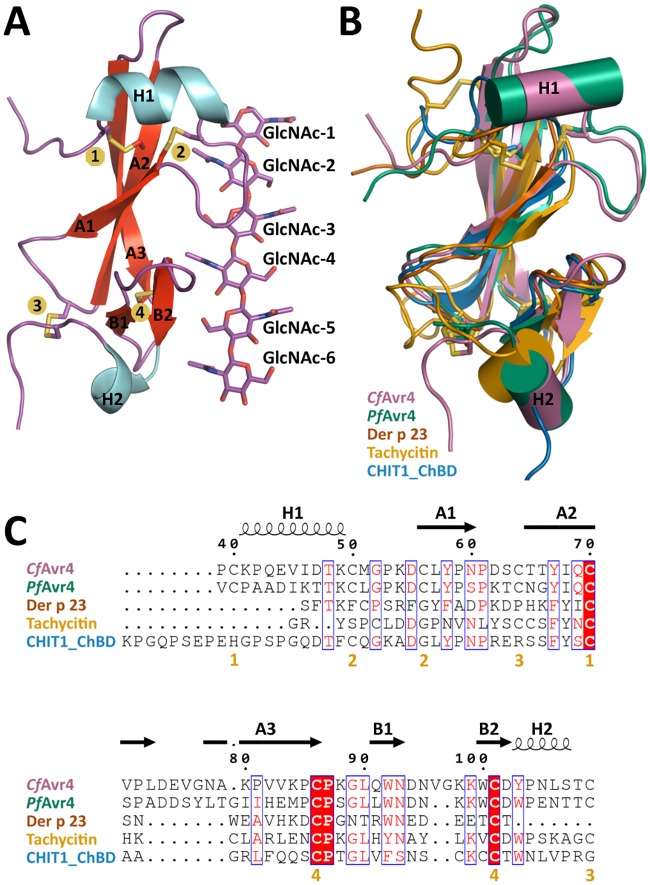
Fig 1. The tertiary and secondary structure of CfAvr4.
(A) Shown is a CfAvr4 monomer bound to the corresponding (GlcNAc)6 molecule (sticks). GlcNAc-1 represents the reducing end of the hexasaccharide sugar. The secondary structure elements are labeled as well as the four disulfide bonds (numbers highlighted in yellow). (B) The five members of CBM14 with known structures are superimposed. CfAvr4 (PDB Id: 6BN0) is shown in purple, PfAvr4 (PDB Id: 4Z4A) in green, Der p 23 (PDB Id: 4ZCE) in brown, tachycitin (PDB Id: 1DQC) in yellow, and ChBDCHIT1 (PDB Id: 5HBF) in blue. (C) Sequence alignment of the five CBM14 family members with known structures. The structural elements of CfAvr4 are shown. Mostly conserved or similar residues are shown in red with the blue boxes. Completely conserved residues are highlighted in red boxes. The disulfide bonds of CfAvr4 are signified by the yellow numbers under the sequences.

