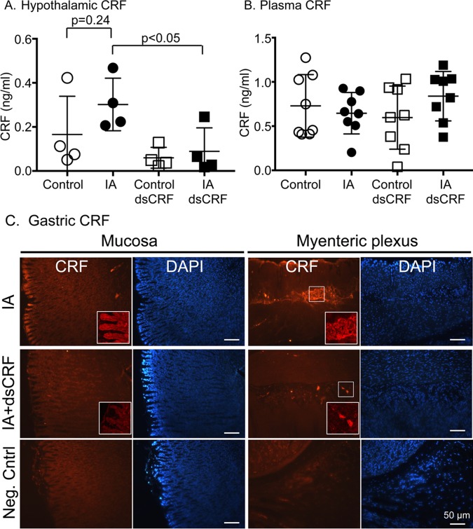
Fig 5. Gastric CRF modulated hypothalamic CRF levels.
(A) CRF levels in the hypothalamus decreased after transient silencing of CRF in the stomach using RNAi in IA-treated rats (p<0.05; Student’s t-tests; IA dsCRF: 0.09 ± 0.05 vs. IA: 0.30 ± 0.06; n = 8/group) compared to control rats, reflecting a bidirectional feedback of the gut-brain axis. (B) Plasma CRF levels were not changed by either IA treatment or after transient silencing of CRF in the stomach. (C) Representative images of CRF-stained stomach sections of IA rats without (top row) and with (middle row) dsCRF treatment (scale bar = 50μm). Bottom row is negative control staining where primary antibody was excluded. Inset: Higher power (40x) images of the brush border of the mucosal layer and the myenteric neurons.

