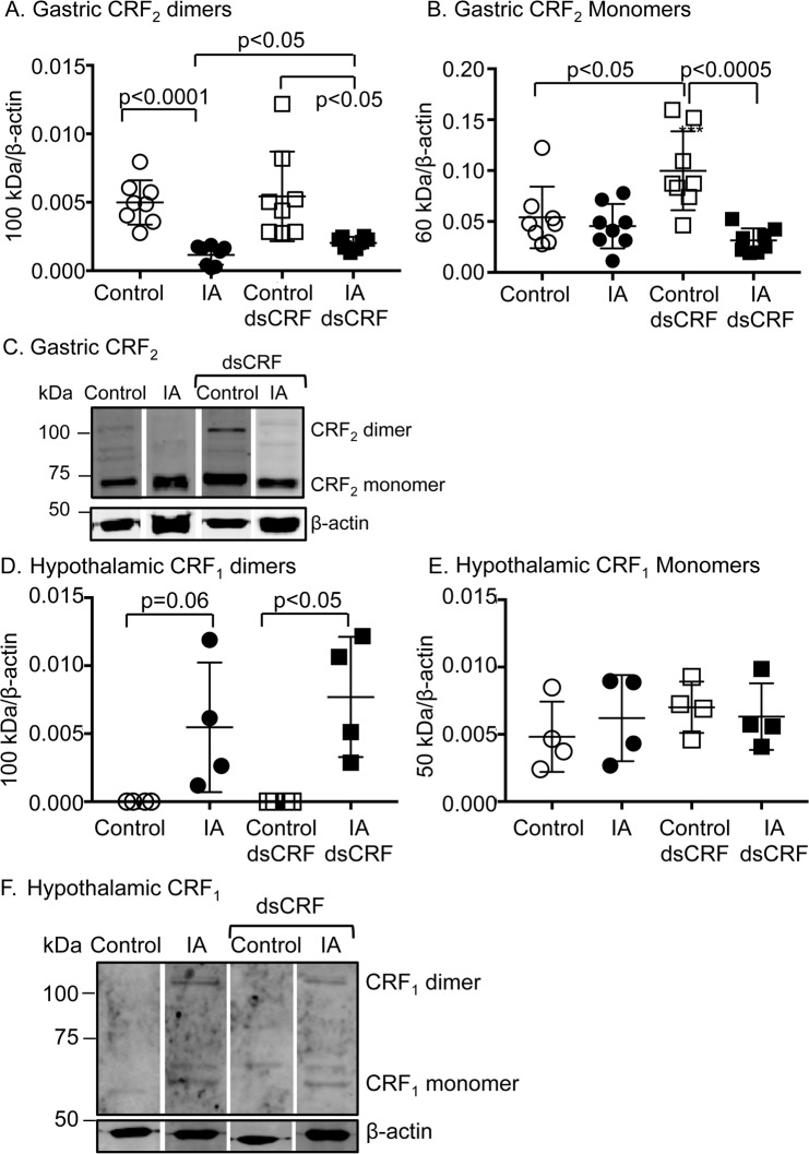
Fig 6. Modulation of CRF receptor levels in FD.
Western blot analyses showing quantification of CRF receptor dimeric and monomeric bands in stomach and hypothalamic protein lysates. β-actin, a housekeeping gene was used as a loading control and its levels were used for normalization. (A) Two-way ANOVA showed significant effect of IA treatment on CRF2 expression levels in the stomach (p<0.0001). CRF2 dimer levels (~100 kDa band) decreased by ~76.8% in IA-treated rats compared to controls (p<0.0001; Student’s t-tests; n = 8/group). CRF-RNAi increased CRF2 dimer expression in FD rats (p<0.05; Student’s t-tests; n = 8/group), but not in control rats. (B) Two-way ANOVA revealed significant main effect of IA treatment and interaction between IA treatment and CRF-RNAi injection (p<0.001 and p<0.005, respectively). CRF2 monomer (~60 kDa band) expression increased in control rats after CRF-RNAi (p<0.05; Student’s t-test; n = 8/group), suggesting that ligand levels regulate receptor expression levels. (C) Representative Western blots showing gastric CRF2 mono- and dimers. (D) Two-way ANOVA revealed significant main effect of IA treatment on CRF1R levels in the hypothalamus (p<0.005). CRF1 dimer (~100 kDa band) expression increased in IA-treated rats compared to control- rats (p = 0.06; Student’s t-tests; n = 4/group). CRF-RNAi further increased CRF1 dimer levels in IA rats (p<0.05; Student’s t-tests; n = 4/group). (E) CRF1 monomer levels (~50 kDa band) in the hypothalamus were not affected by either IA or CRF-RNAi treatments. (F) Representative Western blots showing hypothalamic CRF1 monomers and dimers.

