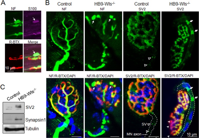
Figure 6. Clustered synaptic vesicles in HB9-Wls-/- axons.
(A) Immunostaining of axonal terminal swelling in gastrocnemius of P21 HB9-Wls-/- mice with anti-NF (Green), anti-S100 (Purple), R-BTX (Red). (B) Immunostaining of NMJs in gastrocnemius of 2-month-old control and mutant mice with anti-NF or anti-SV2 to label synaptic vesicles (Green). R-BTX indicates the AChR (Red). Cell nucleus is labeled with DAPI (Blue). Dashline indicates motor axon. Notice that in the mutant NMJ, there are aberrant deposits of synaptic vesicles in the motor axon (bold white arrow in mutant and empty arrowhead in control), and reduced overlap between pre and post-synapse (white arrowhead). Nucleuses around motor axons are from Schwann cells. (C) Immunoblot of SV2 and Synapsin 1 in phrenic nerve at 2-month-old control and mutant mice.

