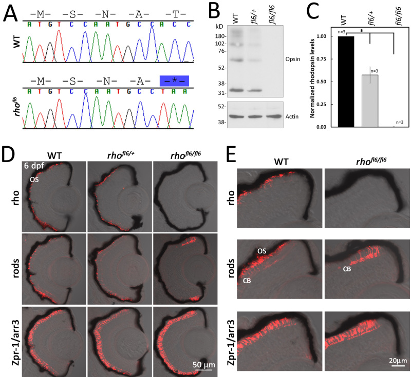
Figure 2.
DNA sequence and histology of rhofl6 encoding a premature stop codon p.17T*. A: Chromatograms overlaid with amino acid sequences comparing WT and rhofl6 allele encoding p.17T*. B: Representative immunoblot analysis of detergent-soluble extracts of retinas from WT, heterozygous rhofl6/+, or homozygous rhofl6/fl6 adults separated by SDS–PAGE, transferred to nitrocellulose, and probed with the 1D1 mab against Rho or mab AC-74 against B-Actin. Immunolabeling of opsin observed as the monomer at 32 kDa and multimers at 64, and 98 kDa were reduced in the heterozygous rhofl6/+ sample and not detected in the homozygous rhofl6/fl6 retina. C: Bar graph of normalized densitometry of immunoblot labeling for Rho (n = 3 animals per genotype), * p<0.025, Kruskal-Wallis test. D: Confocal images of cryosections of retinas from 6 dpf WT, heterozygous rhofl6/+, or homozygous rhofl6/fl6 mutants labeled with antibodies to Rho (1D1, red), rods (4C12, red), or Zpr-1/Arr3a, a selective marker expressed by red/green cones (Zpr-1, red) overlaid with bright-field microscopy. Note the lack of labeling for rods in the central retina and differential labeling for Rho/1D1 and rods/4C12 near the retinal margin of rhofl6/fl6 homozygous larvae, suggesting that rhofl6 is a null allele. E: Higher magnification images of immunolabeling of the dorsal retinal margin WT and homozygous rhofl6/fl6 mutants showing the lack of immunolabeling for Rho, an absence of outer segments (OS), but labeling of the cell bodies (CB) by 4C12 in the homozygous mutant larvae.

