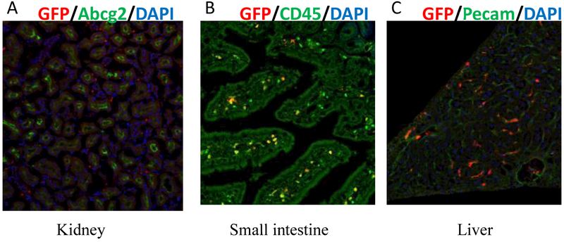
Fig 3. Lack of marking in kidney proximal tubules, hepatocytes and small intestine epithelial cells.
Abcg2CreERT2RosaEYFP mice were treated with a single injection of 4-OHT at E7.5. Fetuses were allowed to be born and grew to adult and tissue sections were stained with anti-GFP antibody, anti Abcg2 antibody Bxp-53 and DAPI. Kidney (A), small intestine (B), liver (C) all at 20X magnification.

